
Faculty Research Highlights
For detailed information about ISMSM Faculty Research please visit the Faculty Research Page.
Structure of Oobleck
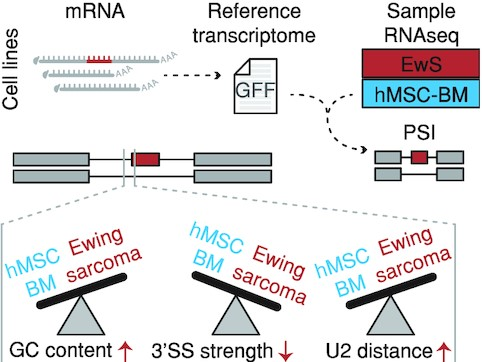
mRNA splicing in EwS
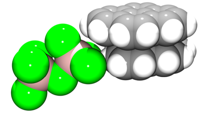
Uneven Charge Distribution
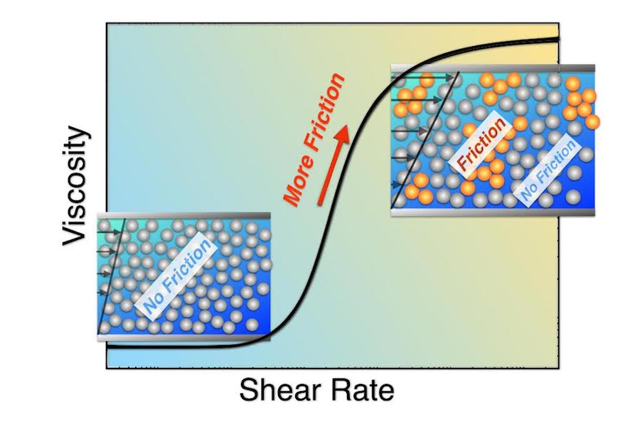
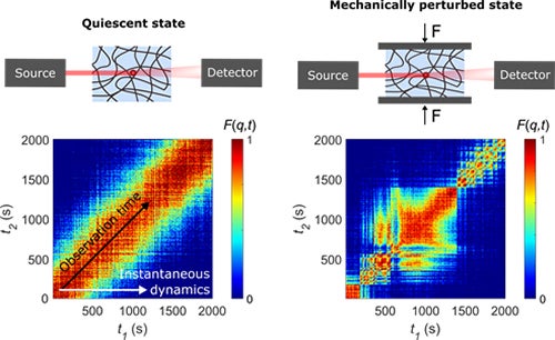
Shear Thickening in Colloidal Suspensions
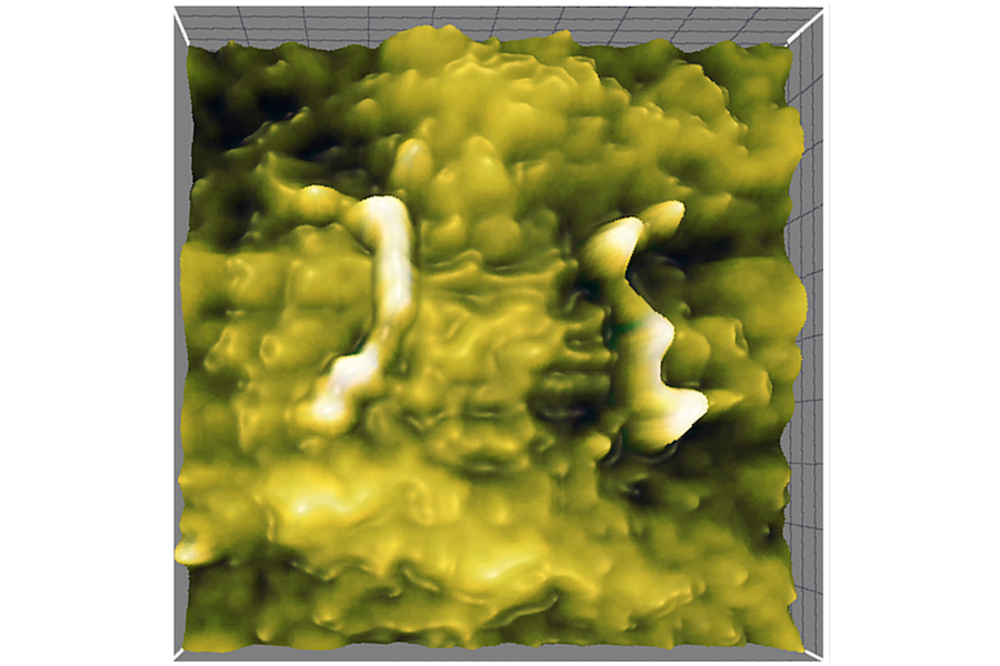
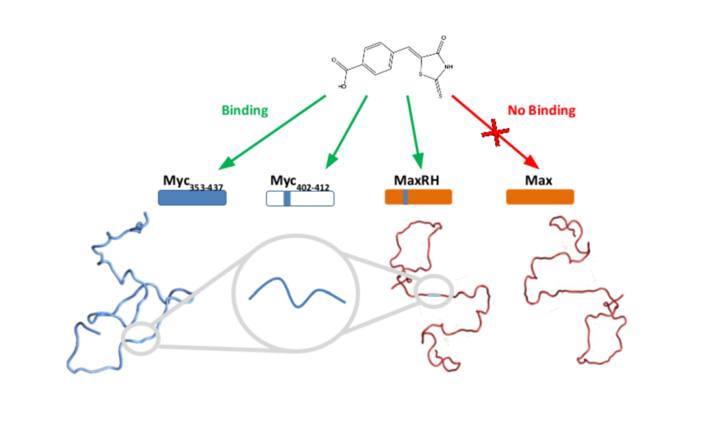
Individual and sub-protein assembly on nanoscale polymeric surfaces different adsorption environments.
Proteins on Surfaces
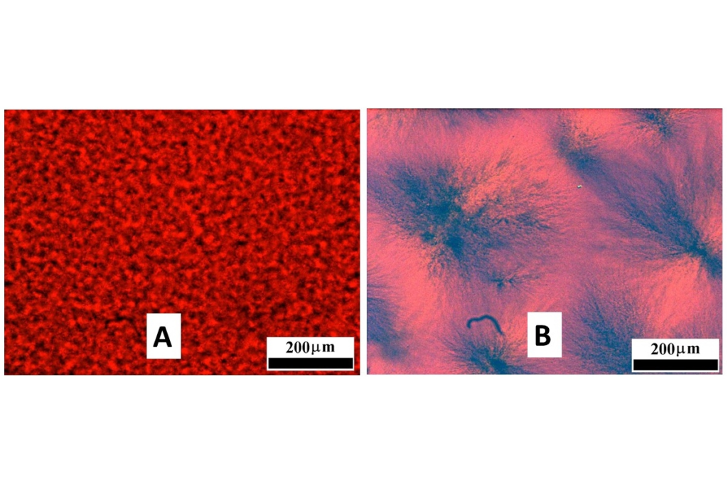
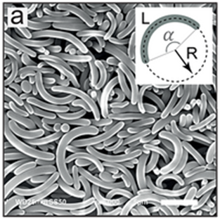
of a hydroxystearamide/silicone mixture formed by (a) fast or (b) slow cooling.
Oranogels
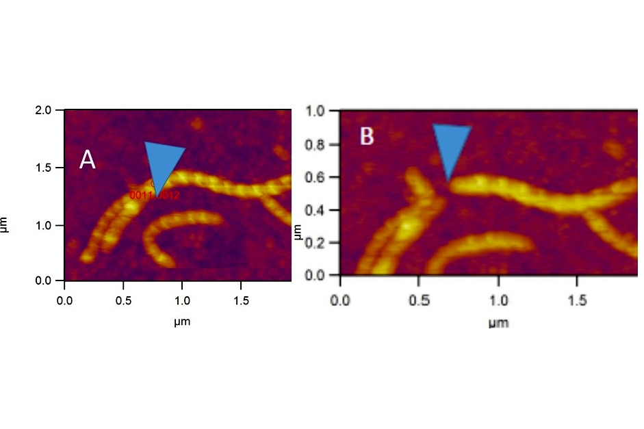
AFM images of a hydroxystearamide/silicone gel before (A) and after (B) a helical fiber aggregate was cut and moved by the AFM tip.
Oranogels
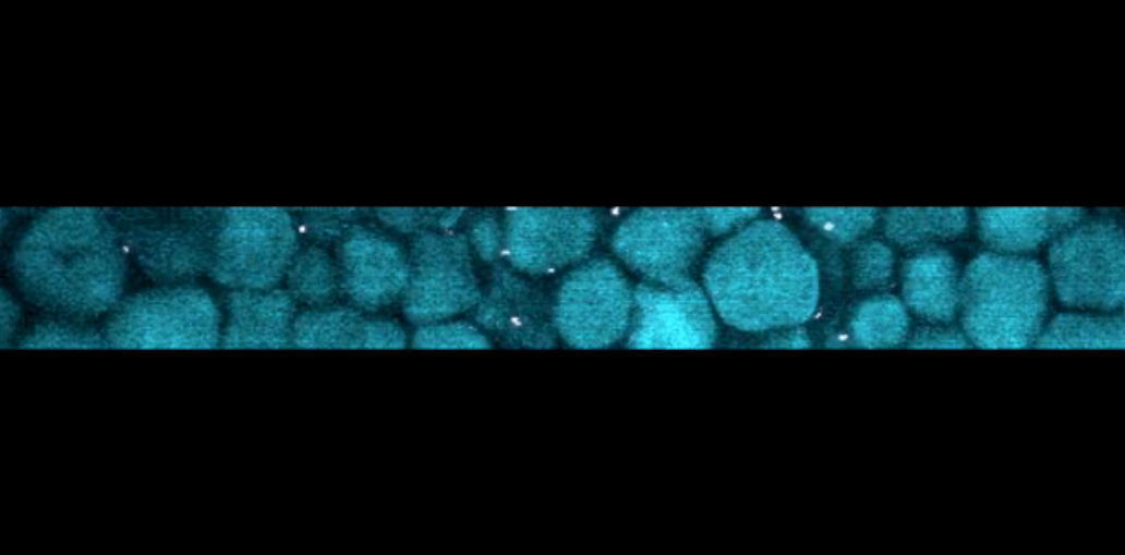
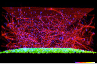
A silkworm surrounded by spun silk, and a confocal image of shear-induced silk fibers.
Confocal microscopy of silk proteins
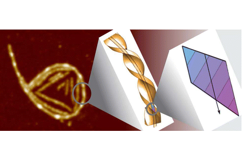
A model polar chiral filament (center, right), by symmetry, develops a spontaneous curvature at the air/water interface.
Helical protein fibers on interfaces
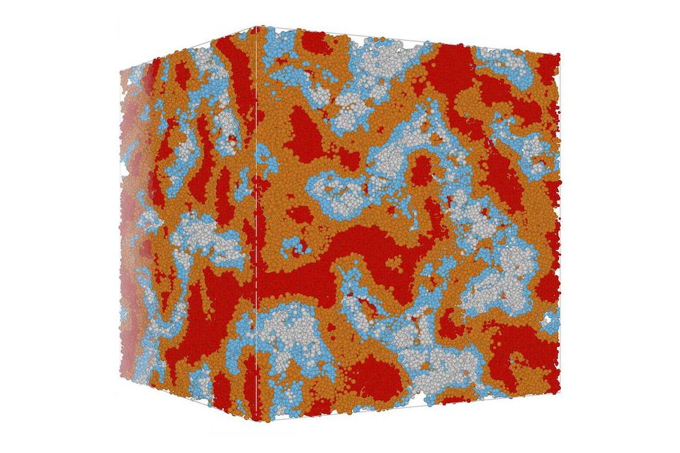
Simulations of Cement Hydration
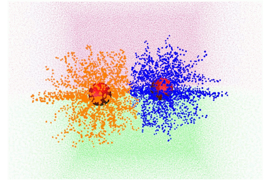
Conformations of core-shell nanoparticles at a liquid interfaces from numerical simulations.
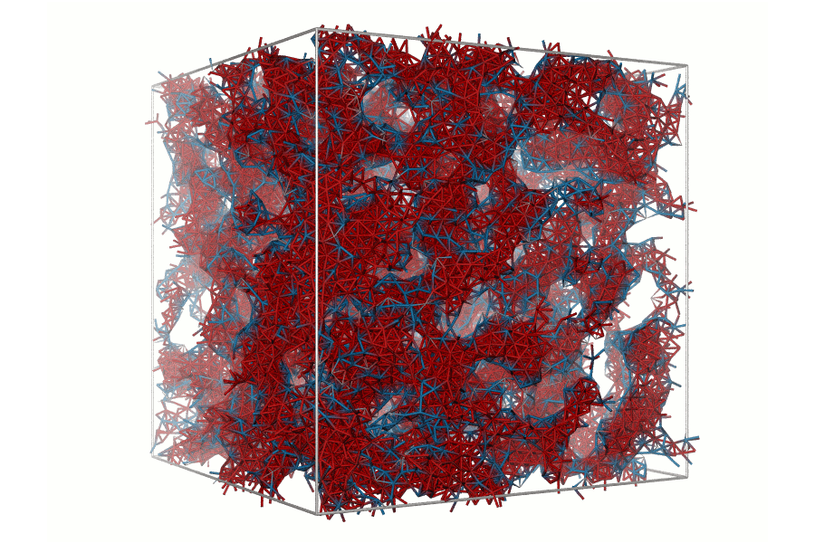
Model of calcium-silicate-hydrate gels formed during cement hydration from numerical simulations.
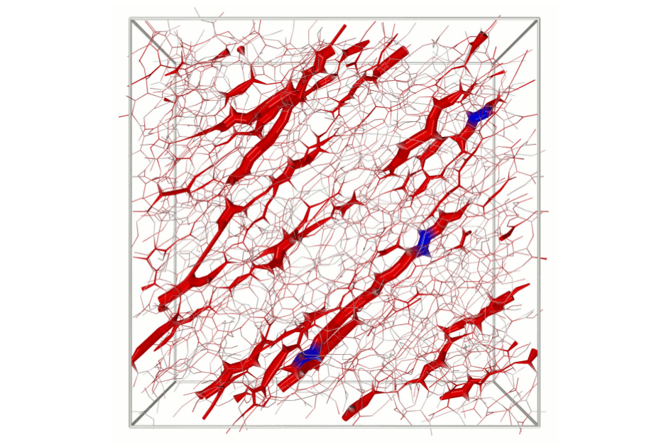
Stress localization in a model colloidal gel under shear.
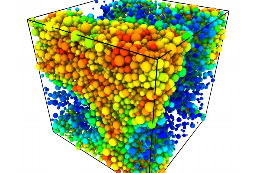
Computer simulation of phase separation in a highly polydisperse colloid.
polydisperse colloidal crystallization
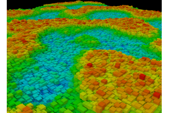
Computer simulation of phase separation in a lipid bilayer, which is thought to be related to the functioning of the cell membrane
lipid membrane phase separation
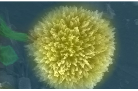
A colorized SEM image of a nanocrystal of tetracyanoquinodimethane.
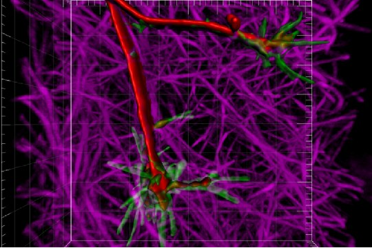
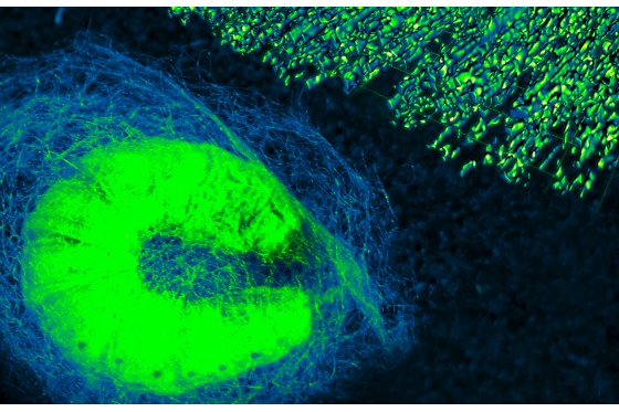
Axon navigating in a 3D collagen gel. The actin from the axon is labeled in green, the microtubules red, and the collagen fibrils in magenta.
Confocal microscopy of axon growth
Boundary stress microscopy of collagen networks
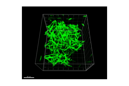
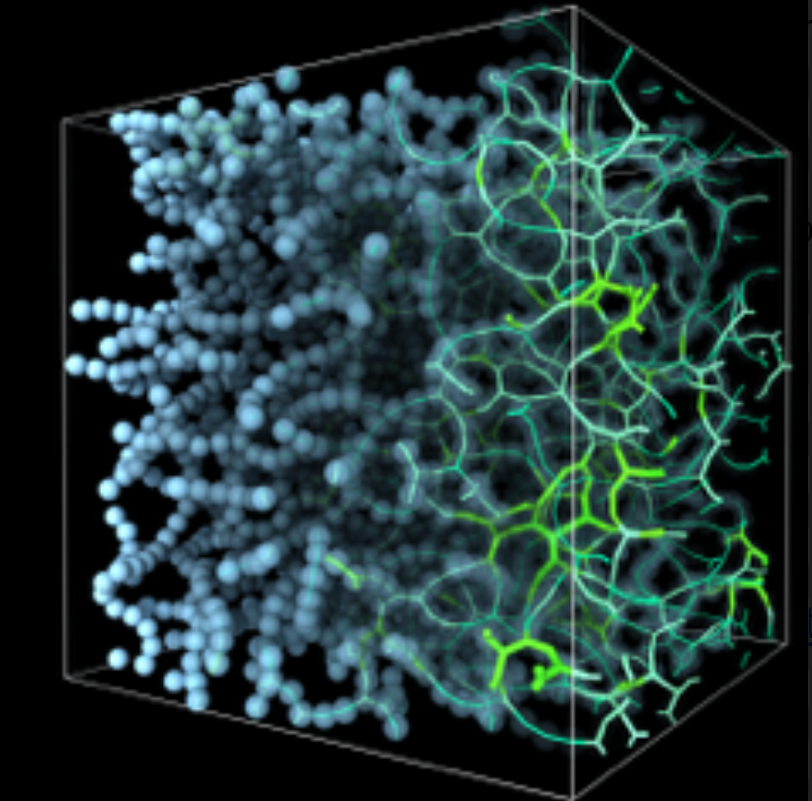
3D confocal microscopy image a disordered aggregate formed by shearing a suspension of µm-scale rods.
Aggregation of rod-like colloids